
“We would like to extend our heartfelt thanks to the investigation site team, especially the investigators and proctors, for their invaluable contributions to this groundbreaking first-in-human trial. Their efforts have been crucial in enabling the publication of these important validating results,” said Peter Hinchliffe, CEO of MedLumics. “As we move forward, we are excited to announce our Series B funding round of €15 million, which will help us achieve our regulatory clinical trial readiness milestone. With these promising results, we are poised to advance toward treating the 100 million patients worldwide affected by Atrial Fibrillation.”
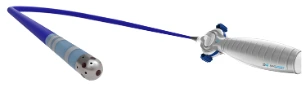
MedLumics® has made a significant breakthrough in the treatment of Atrial Fibrillation (AF) with the development of its AblaView® system, which enables real-time visualization during AF ablation treatments. This innovation is set to improve the precision, safety, and effectiveness of cardiac procedures by addressing key challenges faced by electrophysiologists. The AblaView® system utilizes advanced optical coherence reflectometry (OCR) to visualize critical factors during AF ablation and provide insights that were previously unattainable in real time. The system’s fiber-optic technology offers several unique capabilities that can enhance patient outcomes during the treatment of AF.
The results of a three-month follow-up on the use of the AblaView® system have shown promising outcomes:
The results of this first-in-human study were presented at the AF Symposium in January and later published in Europace in February 2025. Several leading experts in the field of electrophysiology have expressed their enthusiasm about the potential of this technology.
Dr. Atul Verma, from the Electrophysiology Unit, Cardiology Department at McGill University, Montreal, Quebec, Canada, commented on the study, saying, “This first-in-human study demonstrates the ability of optics to determine durable lesions created by PFA. These optical sensors can be adopted to any catheter platform, and we will be moving forward with a full approval trial as the next step.”
Professor Raphael Martins, from the Cardiology Department at CHU Rennes, France, also praised the technology, stating, “It has been a privilege to take part in this first-in-human study. The ability to assess lesion durability in real-time using optical imaging may transform our approach to AF ablation. This represents a significant advancement toward more precise, safer, and more effective treatments for our patients.”
In addition, Professor Sabine Ernst, MD, PhD, FESC, Consultant Cardiologist at the Royal Brompton and Harefield NHS Foundation Trust in London, expressed her excitement on LinkedIn, commenting, “Assessing the ablation lesion in real time: finally, the ‘holy grail’ in electrophysiology is discovered! A new era has begun!”
Founded in 2014, MedLumics S.L. is an ISO 13485-certified medical device company focused on developing optically guided medical devices. The company’s proprietary integrated optics platform combines optical and electrical components, allowing for high-quality real-time imaging during cardiac ablation procedures. MedLumics is supported by various prestigious public and private funds, including an undisclosed corporate partner, and continues to drive innovation in the field of cardiac care.
The company’s commitment to advancing electrophysiology through optical technologies has the potential to revolutionize the way AF ablation is performed, enhancing safety, precision, and patient outcomes in this critical area of heart care.